Description
+ Include: 43 videos + 2 pdfs, size: 8.41 GB
+ Target Audience: pathologists needing to improve their understanding and confidence in the diagnosis of cytopathology specimens
+ Sample video: contact me for sample video
+ Information:
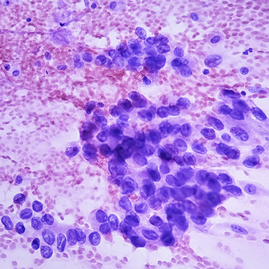
Cytopathology to Refine Diagnostic Skills
Take your diagnostic skills to the next level with this engaging, practice-focused online course. Cytopathology — part of our Masters of Pathology Series — reviews the latest updates to WHO reporting systems and explores how molecular testing and other ancillary studies are transforming the way small biopsy and cytopathology specimens are assessed.
Geared towards those who may not have formal training in cytopathology, the succinct, high-yield lectures in this continuing medical education program are divided by specimen type and will help you to:
- Gain a deeper understanding of how The Bethesda System for Reporting Thyroid Cytopathology has reduced unnecessary surgical interventions
- Consistently generate standardized reports that are more useful and better understood by clinicians
- Avoid common pitfalls like overinterpreting benign findings or relying too heavily on surgical pathology when results are discordant
- Confidently identify benign and reactive findings in cytopathology specimens
- And more…
Date of Original Release: January 15, 2026
Learning Objectives
At the completion of this course, you should be able to:
- List ancillary tests that should be ordered on lung specimens when a new diagnosis of non-small cell carcinoma is made
- Describe changes made in the newest version of The Bethesda System for Reporting Thyroid Cytopathology
- Name the diagnostic categories used in The Paris System for Reporting Urinary Cytology
- Understand situations in which a diagnosis of Salivary Gland of Uncertain Malignant Potential (SUMP) can be made
- Recognize the cytomorphologic features of common bone and soft tissue tumors on fine needle aspiration
- Compare different methodologies used for human papillomavirus (HPV) testing
Intended Audience
Pathologists needing to improve their understanding and confidence in the diagnosis of cytopathology specimens.
+ Topics:
The Bethesda System for Reporting Thyroid Cytopathology Third Edition – Paul A. VanderLaan, MD, PhD
Molecular Testing in Thyroid FNA Specimens – Michiya Nishino, MD, PhD
Thyroid Cytomorphology Part I – Christopher J. VandenBussche, MD, PhD
Thyroid Cytomorphology Part II – Christopher J. VandenBussche, MD, PhD
Instructive Thyroid FNA Cases – Christopher J. VandenBussche, MD, PhD
The Milan System for Reporting Salivary Gland Cytopathology Second Edition – William C. Faquin, MD, PhD
Instructive Salivary Gland FNA Cases – Zahra Maleki, MD
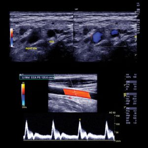
FNA of Lateral Neck Lesions – A Case-Based Approach – Darcy A. Kerr, MD
Approach to Liver Cytopathology and Small Biopsies – M. Lisa Zhang, MD
Update on the WHO Reporting System for Pancreaticobiliary Cytopathology – Martha Bishop Pitman, MD
FNA Cytology of Solid Pancreatic Lesions – Monica T. Garcia-Buitrago, MD, FCAP
Approach to Pancreatic Cyst Cytology – M. Lisa Zhang, MD
Instructive Pancreas FNA Cases – Christopher J. VandenBussche, MD, PhD
Serous Cavity I – The International System for Serous Fluid Cytopathology – Ashish Chandra, FRCPath, DipRCPath (Cytol)
Serous Cavity II – Pleural Fluid – Malignant Tumors and Mimickers – Peter Illei, MD
Serous Cavity III – Pelvic Washing Specimens – Christopher J. VandenBussche, MD, PhD
Serous Cavity IV – Challenges Cases – Pelvic Washing Specimens – Christopher J. VandenBussche, MD, PhD
Bone and Soft Tissue (BST) I – The WHO Reporting System for Soft Tissue Cytopathology – Ivan Chebib, MD, FRCPC
Bone and Soft Tissue (BST) II – A Pattern-Based Approach – Vickie Y. Jo, MD
Bone and Soft Tissue (BST) III – An Update on Immunohistochemical Markers – Vickie Y. Jo, MD
Cytopathology of Bone Tumors – Valerie A. Fitzhugh, MD
Cytopathology of Soft Tissue Tumors – Valerie A. Fitzhugh, MD
The International System for Reporting Lung Cytopathology – Fernando Schmitt, MD, PhD, FIAC
Pulmonary I – Cytology of NSCLC – Does Morphology Matter? – Peter Illei, MD
Pulmonary II – Lung Tumors other than NSCLC – Peter Illei, MD
Pulmonary III – Ancillary Testing in NSCLC: Why, When, and How to Perform? – Peter Illei, MD
Pulmonary IV – Non-Neoplastic Pulmonary Cytology: Bugs, BALs, and Beyond – Peter Illei, MD
Kidney I – Fine Needle Aspiration and Small Biopsy Specimens – Madelyn Lew, MD
Kidney II – Fine Needle Aspiration and Small Biopsy Specimens – Madelyn Lew, MD
Breast I – Introduction to the Yokohama System – Gary Tse, MBBS
Breast II – Challenging Cases in Breast Cytology – Christine Noga Booth MD
Urine I – The Paris System for Reporting Urinary Cytology – Christopher J. VandenBussche, MD PhD
Urine II – Benign and Reactive Findings in Urine Specimens – Christopher J. VandenBussche, MD PhD
Urine III – Neoplastic Findings in Urine Specimens – Christopher J. VandenBussche, MD PhD
Urine IV – Instructive Urine Cytology Cases – Madelyn Lew, MD
Miscellaneous FNA Challenge Cases – Christopher J. VandenBussche, MD PhD
HPV Biology and Testing in Pap Test Specimens – Michael J. Thrall, MD
ASCCP Guidelines and the Role of Cytology in Screening – Michael J. Thrall, MD
Pap Test Cytomorphology I – Squamous Lesions – Donna K. Russell, MEd, CT(ASCP)HT, CFIAC
Pap Test Cytomorphology II – Glandular and Rare Lesions – Donna K. Russell, MEd, CT(ASCP)HT, CFIAC
Instructive Pap Test Cases – Donna K. Russell, MEd, CT(ASCP)HT, CFIAC
Cerebrospinal Fluid and Vitreous Fluid Specimens – Cheng-Ying Ho, MD, PhD
Lymph Node Cytology – A Case Based Approach in Small Biopsies of Lymph Nodes – Sara E. Monaco, MD


Reviews
There are no reviews yet.