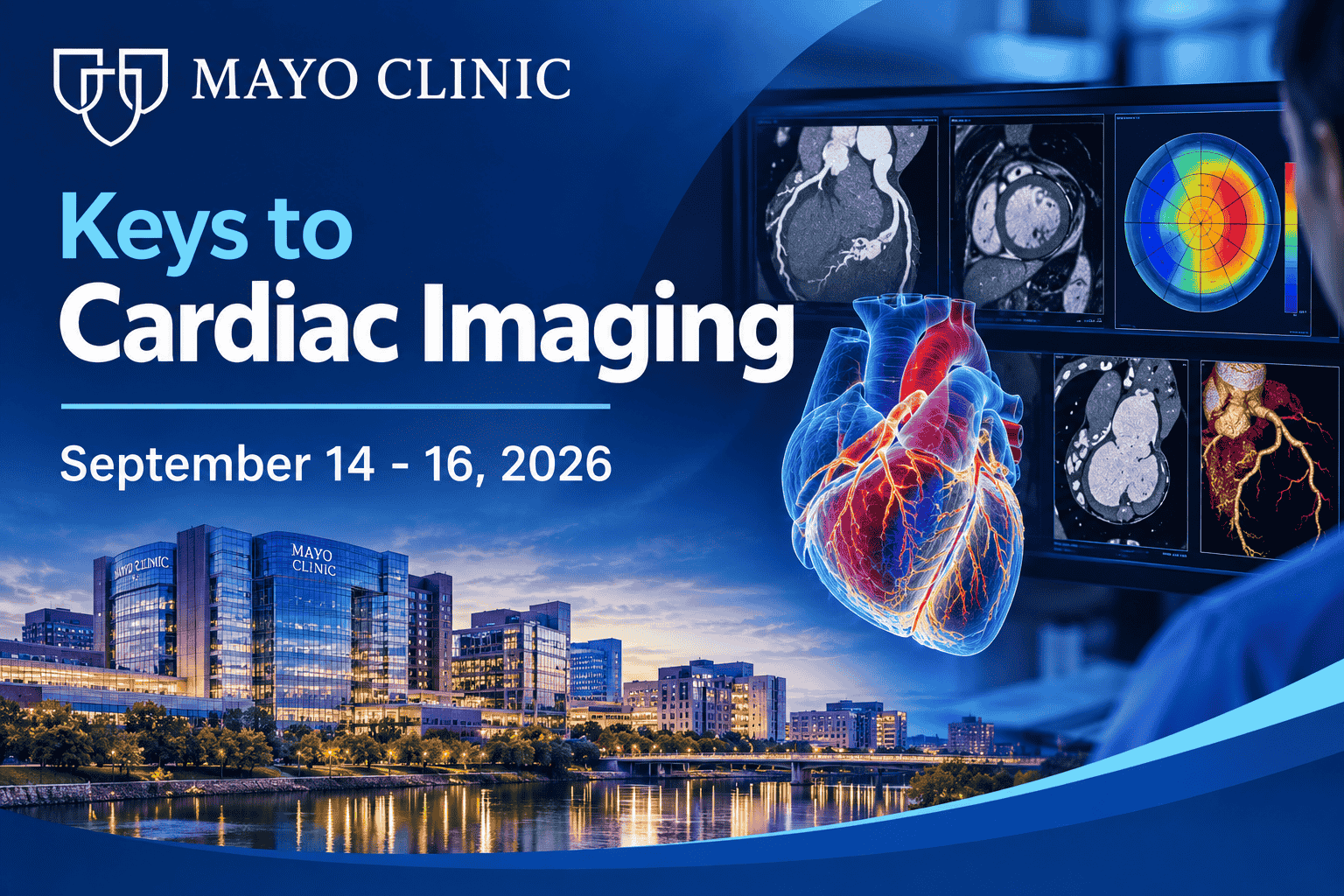
Description
+ Include: videos + file sub vtt + pdf, size: GB
+ Target Audience: cardiologists, radiologists, cardiac anesthesiologists, general internists, internal medicine subspecialists, sonographers, radiology technologists
+ Sample video: contact me for sample video
+ Information:
COURSE LOCATION
The Biltmore Miami-Coral Gables 1200 Anastasia Avenue Coral Gables, FL 33134
This course will explore basic and cutting-edge imaging techniques to improve patient care and hone individual skills in performance and interpretation of echocardiography, nuclear cardiology, cardiac computed tomography, and cardiovascular magnetic resonance imaging. Led by a distinguished faculty of experts, the course will highlight recent advances, practical applications, and limitations of each imaging technique. Educational sessions will focus on the clinical utility of imaging in the diagnosis, risk stratification, and management of a wide range of cardiovascular conditions. Key topics will include valvular heart disease, coronary artery disease, cardiomyopathies, and functional assessment. Through this course, clinicians will obtain a greater understanding of the individual and complimentary roles of the wide range of imaging modalities in providing optimal care patients with cardiovascular diseases.
LEARNING OBJECTIVES
Upon completion of this activity, participants should be able to:
- develop skills to select appropriate multimodality cardiac imaging;
- utilize cutting edge techniques for advanced imaging for cardiovascular disease;
- utilize diagnostic and clinical decision-making skills in the care of cardiovascular patients using multimodality cardiac imaging; and,
- identify appropriate use of imaging indications, contraindications, and rationale for testing.
INTENDED AUDIENCE
This course is designed for the cardiologists, radiologists, cardiac anesthesiologists, general internists, internal medicine subspecialists, sonographers, radiology technologists, nurse practitioners, physician associates, and other primary healthcare providers interested in appropriate imaging for cardiovascular disease assessment and diagnosis.
+ Topics:
*Note: these are continuous video recordings during the conference, they include individual lectures mentioned in the Detail section below
MONDAY, SEPTEMBER 14, 2026
Welcome and Course Overview
LATEST ADVANCES IN CARDIOVASCULAR IMAGING: ECHO, CT, PET, AND CMR
Photon-Counting Cardiac CT: The Next Generation of Coronary Imaging
Beyond Perfusion: Molecular Imaging of Cardiovascular Disease
Contemporary Cardiovascular MRI: Techniques, Applications, and Future Directions
Artificial Intelligence and Wearable Technologies in Echocardiography
Artificial Intelligence in Cardiovascular Imaging: Transforming Image Interpretation and Clinical Decision Making
ISCHEMIC HEART DISEASE
Coronary CT for Calcium Score: Risk Stratification and Clinical Integration
Nuclear Stress Testing, Including SPECT and PET
Cardiac MRI for Stress Testing and Viability
Coronary CTA: Native Coronary Arteries, Bypass Grafts, and Flow by FFR
Plaque Characterization and CT Evaluation for PCI
ILLUSTRATIVE CASES: BEST IMAGING CASES OF THE YEAR
Case 1
Case 2
HANDS-ON ECHO WORKSHOP: POINT OF CARE ULTRASOUND (POCUS) (NOT INCLUDE LIVESTREAMS)
This hands-on workshop provides a practical introduction to Cardiac Point-of-Care Ultrasound (POCUS) for rapid bedside cardiovascular assessment. Participants will learn focused cardiac imaging techniques, image acquisition, and interpretation to aid in the evaluation of ventricular function, volume status, and hemodynamic instability. In-Person Only Attendance – this session is not available via livestream.
Cardiac POCUS: Assessing the LV – Basic Views, Wall Motion, EF
Hands-On Echo Scanning with Advanced Cardiac Sonographer
Cardiac POCUS: Assessing the RV, Pulmonary Pressures, and IVC
Hands-On Echo Scanning with Advanced Cardiac Sonographer
Cardiac POCUS: Assessment of Valvular Heart Disease
Hands-On Echo Scanning with Advanced Cardiac Sonographer
TUESDAY, SEPTEMBER 15, 2026
Livestream airs in Eastern Daylight Time
STRUCTURAL / INTERVENTIONAL HEART DISEASE I
Echo Evaluation of Aortic Stenosis
TAVR CT: Aortic Valve Calcium Score, Annular Measurements, Peripheral Vessel Assessment
TAVR CT: Beyond the Basics: Valve-in-Valve, Bicuspid Aortic Valve, and Alternate Access Assessment
Severe Mital Valve Stenosis: Diagnosis and Management
Predicting the Perfect Seal: Echo, CT, and AI Simulation for LAA Closure Planning
STRUCTURAL / INTERVENTIONAL HEART DISEASE II
Tricuspid Valve: A Case-Based Multimodality Approach to Grading and Intervention Planning
CTEPH: Identifying Candidates for Balloon Pulmonary Angioplasty in the Modern Era
Mitral Annular Disjunction, Scar, and Arrhythmias
Multimodality Evaluation of Endocarditis
4-D Flow in Structural Disease
SPECIAL TOPICS
Multimodality Imaging in the Evaluation of Cardiac Amyloidosis
Incidental Noncardiac Findings—What Do I Do With This?
3-D Modeling: Medical Applications Using Imaging Data
OPTIONAL HANDS-ON ECHO WORKSHOP: TRANSESOPHAGEAL AND INTRACARDIAC ECHO (NOT INCLUDE LIVESTREAMS)
Hands-on Simulation of TEE and ICE for Tricuspid and Left Atrial Appendage Interactive hands-on session provides participants with practical, simulation-based experience in the complementary roles of Transesophageal Echocardiography (TEE) and Intracardiac Echocardiography (ICE) in structural heart interventions targeting the tricuspid valve and the left atrial appendage (LAA). Participants will develop a systematic approach to pre-interventional TEE evaluation, including comprehensive anatomical assessment of tricuspid valve morphology and pathology, LAA anatomy, thrombus exclusion, and patient selection criteria. Emphasis will be placed on acquiring and interpreting the key imaging windows, measurements, and parameters that inform procedural planning and device selection prior to intervention. The session incorporates the procedural role of ICE, with participants gaining hands-on experience using intracardiac imaging for real-time guidance during structural interventions. Training will focus on catheter manipulation, transseptal puncture guidance, device positioning, and post-deployment assessment — equipping participants with the skills to navigate complex intraprocedural scenarios with confidence. This session is ideal for cardiologists, interventionalists, echocardiographers, fellows, sonographers, and allied healthcare professionals seeking to build a comprehensive, imaging-guided approach to structural heart disease — from preprocedural workup through procedural execution.
WEDNESDAY, SEPTEMBER 16, 2026
Livestream airs in Eastern Daylight Time
CARDIOMYOPATHIES
Echo Evaluation of Cardiomyopathy Including Strain
Multimodality Imaging of Hypertrophic Cardiomyopathy
CMR Assessment of Myocarditis
Cardiac Myosin Inhibitors in HCM: What the Imager Needs to Know
Arrhythmogenic Cardiomyopathy (ARVC/ACM)
SPECIAL TOPICS
Cardiac MRI Safety
Artifacts in Cardiovascular MRI: How to Identify and (Sometimes) Correct Them
MRI Evaluation of Cardiac Masses
Utility of T1 and T2 Mapping in Cardiomyopathy Assessment
Troubleshooting and Improving Cardiac Imaging Protocols


Reviews
There are no reviews yet.